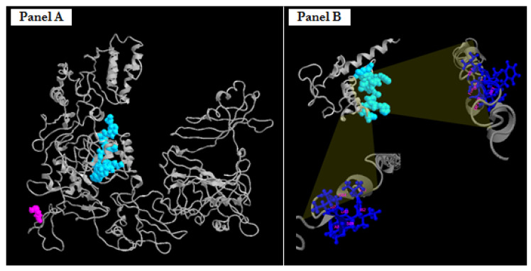
Figure 5.
Predicted three-dimensional structures of FBRSL1, wild-type (panel A) and truncated proteins p.Cys125Leufs*7 (panel B). Highlighted in light blue space-filling mode is the predicted protein domain, which extends from the amino acid residues 81 to 93 capable of binding the amino acid residues of FBRSL1 in the same position. Highlighted in blue ball-and-stick mode are the specific amino acid residues (at the top right, 85, 86, 87, 90, 91, 92, 93 aa; on the bottom left, 81, 82, 83, 84, 87, 88, 89 aa) able to bind the light blue domain.

