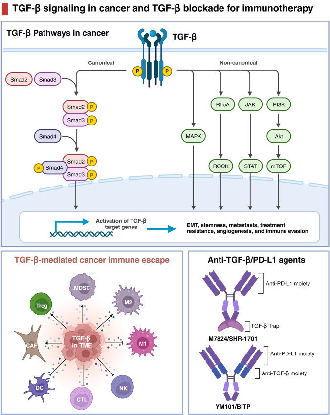
Fig. 7.
TGF-β signaling in cancer and TGF-β blockade for immunotherapy. The top panel illustrates the TGF-β signaling pathway in cancer cells, including the canonical Smad-dependent pathway and the non-canonical pathways involving various intracellular mediators such as MAPK, PI3K/Akt, and mTOR, leading to cellular processes like EMT, stemness, metastasis, treatment resistance, angiogenesis, and immune evasion. The bottom left panel depicts the role of TGF-β in the tumor microenvironment (TME), highlighting its immunosuppressive effects that facilitate cancer immune escape by interacting with various immune cells such as Treg, MDSC, M1/M2 macrophages, DC, NK, and CTL. The bottom right panel presents a schematic representation of innovative anti-TGF-β/PD-L1 therapeutic agents, demonstrating dual blockade strategies, as exemplified by M7824/SHR-1701, which combines a TGF-β trap with an anti-PD-L1 moiety, and YM101/BITP, which features both anti-TGF-β and anti-PD-L1 moieties for enhanced immunotherapy efficacy. Adapted from “Canonical and Non-canonical TGF-β Pathways in EMT”, by BioRender.com (2024). Retrieved from https://app.biorender.com/biorender-templates

