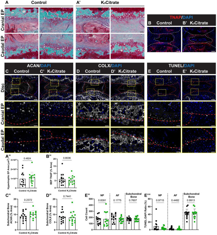
Figure 5. Quantitative histology reveals endplate chondrocytes and a chronic repair response may drive disc mineralization in LG/J mice, without a cellular response to K3Citrate.
(A-A”) Safranin O/Fast Green/Hematoxylin-staining revealed what appeared to be aggregates of hypertrophic chondrocytes in the cartilaginous endplates, and the (B) area of these aggregates did not change with K3Citrate supplementation. Quantitative immunohistological staining subchondral bone/endplate space for hypertrophic chondrocyte markers (C-C”) aggrecan (ACAN) and (D-D”) collagen X (COLX), as well as (E-E”’) TUNEL staining to delineate cell death, provide evidence of lesions along the cartilaginous endplates, resembling fracture healing in bone; this was unattenuated in K3Citrate mice. (Control mice: n=7 mice (2F, 5M); K3Citrate mice: n=7 mice (3F, 4M); 2 discs/mouse, 14 discs/treatment; Ca6-Ca8) Data are shown as mean ± SD. Significance was determined using an unpaired t-test or Mann-Whitney test, as appropriate.

