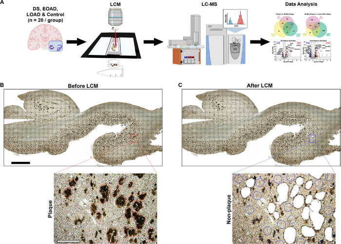
Figure 1.
Schematic of the localized proteomics protocol. A. Laser-capture microdissection of 2 mm2 total area of amyloid-β plaques from hippocampus and adjacent temporal cortex from FFPE autopsy brain tissue from control, DS, EOAD and LOAD (n=20 cases/experimental group). Amyloid plaque proteins were quantified by label-free mass spectrometry and posteriorly analyzed. B-C. Microphotographs of a typical brain tissue section immunolabeled against Aβ illustrate the precise microdissection of amyloid plaques before (B) and after LCM (C). 2 mm (black bar, top) and 200 μm (white bar, bottom).

