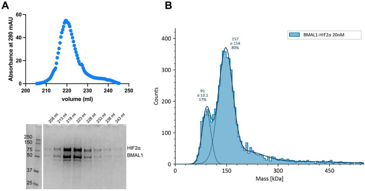
Figure 2: Purified BMAL1 and HIF2α form a stable complex in vitro.
(A) Heparin chromatography elution of BMAL1 and HIF2α co-expressed in insect cells. SDS-PAGE analysis shows a co-eluted stoichiometric complex of BMAL1-HIF2α. (B) Mass photometry of purified BMAL1-HIF2α complex. A minor peak centered at 91 kDa corresponds to the molecular weight of HIF2α, suggesting that it is in slight excess. The major peak, centered at 157 kDa, is consistent with the calculated molecular weight for the BMAL1-HIF2α heterodimer.

