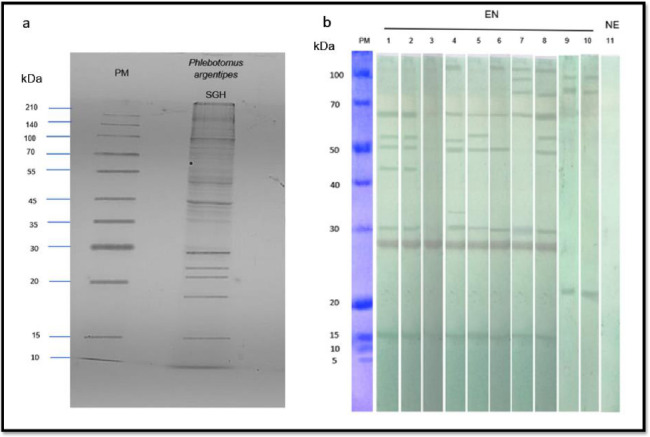
Figure 1. Identification of antigenic Phlebotomus argentipes salivary proteins.
(a) The SGH protein profile of Ph. argentipes. 12% SDS–PAGE gel stained by Coomassie Blue. (b) Western blots showing the reactivity of IgG in endemic sera against Ph. argentipes salivary gland antigens (lane 1–8), or Culex spp. salivary gland antigens (lane 9–10: against sera of the endemic individuals screened in lanes 1 and 8). Lane 11: a non-endemic serum sample screened against Ph. argentipes salivary gland antigens. PM: protein marker; SGH: salivary gland homogenate; kDa: Kilodaltons; EN: endemic sera; NE: Non-endemic sera. The uncropped gel image and blot image can be found as supplementary Fig. S1 and Fig. S2 online.

