
Table 1.
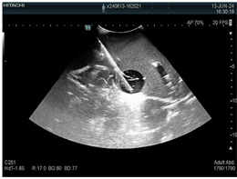
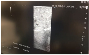
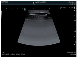
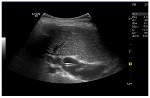
Imaging assessment of gelatin liver phantoms using several imaging devices compared to the same procedures on real patients.
| Examination/ Procedure |
Examination/Procedure on Gelatin Liver Phantom | Examination/Procedure on Real Liver | |
|---|---|---|---|
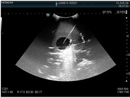
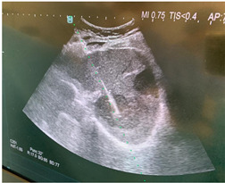
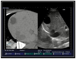
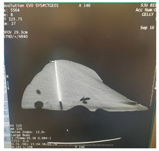
| 1 | US examination |
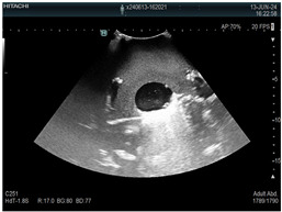 * * |
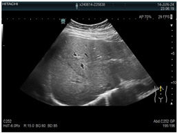 * * |
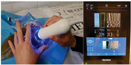
| 2 | Elastography |
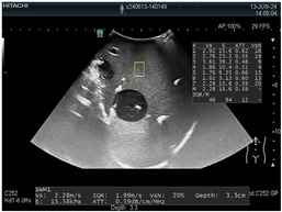 * * |
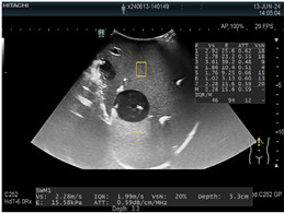 * * |
| 3 | Fibroscan |
 * * |
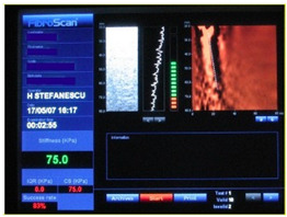 ** ** |
| 4 | US-guided tumor puncture/biopsy |
 * * |
 * * |
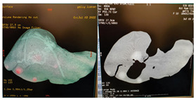
| 5 | RFA needle insertion into the tumor |
 * * |
 * * |
| 6 | US-guided Biliary drainage |
 * * |
 *** *** |
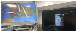
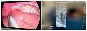
| 7 | Laparoscopic US-guided tumor puncture/biopsy |
 * * |
 * * |
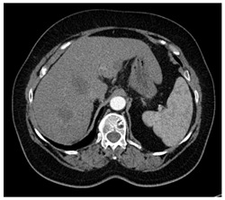
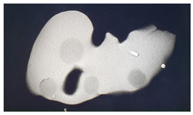
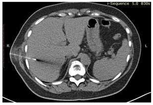
| 8 | CT-scan examination |
 * * |
 4* 4* |
| 9 | MRI examination |
 * * |
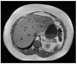 4* 4* |
| 10 | US-CT fusion examination |
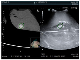 * * |
 5* 5* |
| 11 | CT-guided tumor puncture/biopsy |
 * * |
 6* 6* |
* Personal archive (Dr. Radu Claudiu Elisei). ** Courtesy of Dr. Horia Stefanescu (“Prof. Dr. O. Fodor”, Regional Institute of Gastroenterology and Hepatology, Cluj-Napoca, Romania). *** Courtesy of Dr. Tudor Mocan (“Prof. Dr. O. Fodor”, Regional Institute of Gastroenterology and Hepatology, Cluj-Napoca, Romania). 4* Archive of Bistrita Emergency Clinical County Hospital, Romania. 5* Courtesy of Hiatchi Ltd./Romania. 6* Courtesy of Dr. Andrei Roman (Oncology Institute Cluj-Napoca, Romania).