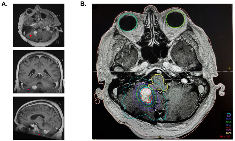
Figure 1.
Stereotaxic radiosurgery planification. (A) Cerebral MRI T1 images prior to stereotactic radiosurgery, with a demonstration of the polylobulated lesion in the axial view, coronal view, and sagittal view. (B) Isodose coverage of the GTV (in red) for the 18 Gy radiosurgery plan. The blue contour is an expansion of 1 mm of the GTV treated at an 87% isodose because of the proximity with the brainstem. A total of 83% of the tumor volume was covered by the 18 Gy isodose, and 99.9% was covered by the 15 Gy isodose.

