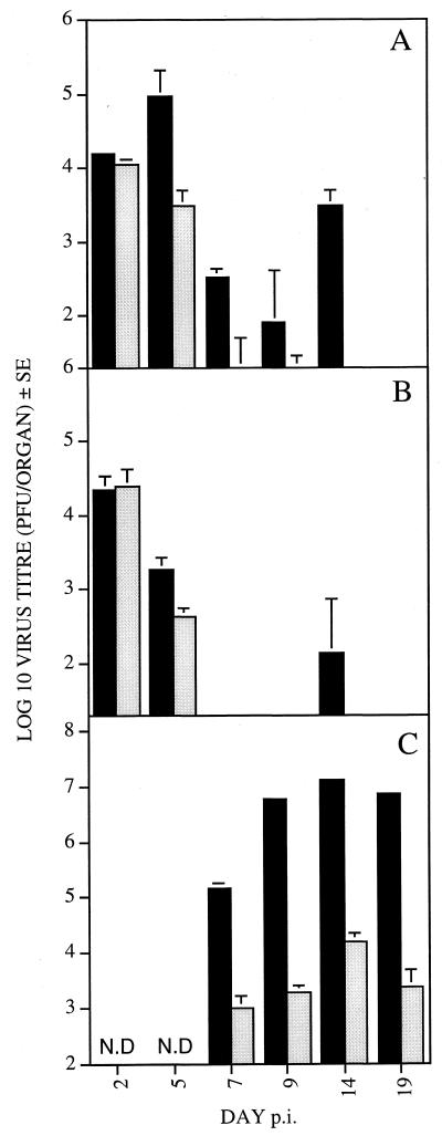
FIG. 6.
Growth of wt (black bars) and Δm131Z (grey bars) virus in the spleens (A), livers (B), and salivary glands (C). Groups of four BALB/c mice were infected i.p. with 104 PFU of the relevant virus, and spleens, livers, and salivary glands were harvested on the days indicated. Organs were homogenized and stored at −80°C before being used for titer determination on MEF. Virus titers are expressed as mean log10 per organ, with standard error. The y axis is cropped to indicate the lower limit of virus detection. N.D, not determined.

