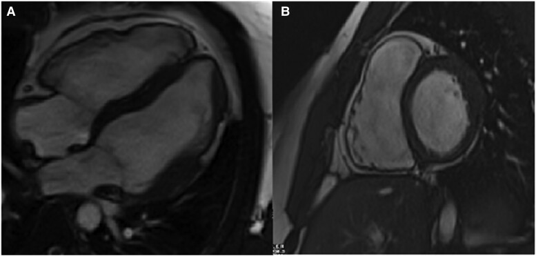
Figure 2.
Cardiac magnetic resonance four-chamber (A) and short-axis (B) images demonstrating right ventricular enlargement compared with the left ventricle. The right ventricular end-diastolic volume was 260 mL and 135 mL/m2 when indexed to the body surface area. The right ventricular ejection fraction was 37%. Focal thickening of the mid-anterior, anterolateral, and inferolateral wall segments with associated transmural delayed gadolinium enhancement and hypokinesis of the left ventricle. Structural abnormalities are characterized by non-ischaemic late gadolinium enhancement affecting the subepicardial and less often the midmyocardial layers. The origin of the fibrofatty infiltration of the ventricular myocardium is thought to be the second heart field cardiac fibroadipocyte progenitor cells. These cells express desmosome genes and have bimodal potential for differentiation into fibrogenic or adipogenic pathways. Increased accumulation of plakoglobin, also known as junction plakoglobin or gamma-catenin, in the nucleus results in the increase in the adipogenic factors and reduction of the inhibitors of adipogenesis (connective tissue growth factor). Adipogenesis of the heart is also mediated by the downregulation of micro-RNA-184 (miR-184). Cine images are presented in Supplementary material online. The use of diagnostic criteria for arrhythmogenic cardiomyopathy is a two-step process. First, one must determine the number of major and minor criteria for both right and left ventricular involvement by applying the multiparametric approach, covering six categories: (i) morpho-functional abnormalities, (ii) structural myocardial abnormalities, (iii) electrocardiogram repolarization abnormalities, (iv) electrocardiogram depolarization abnormalities, (v) ventricular arrhythmias, and (vi) family history and molecular genetics.

