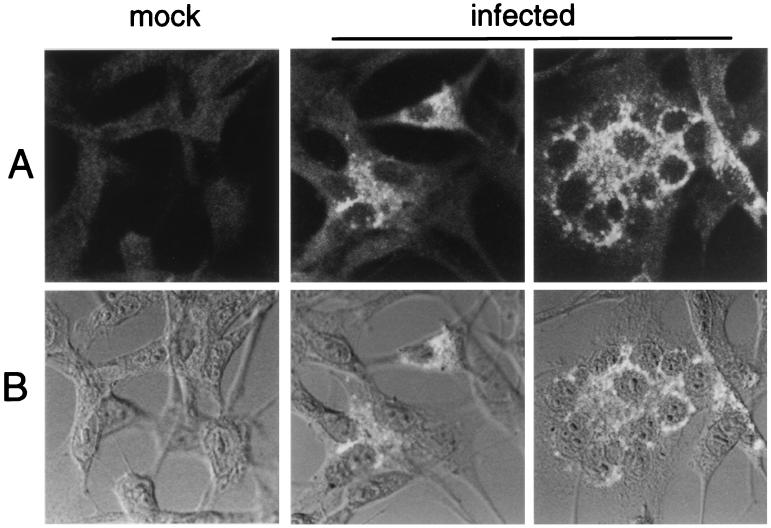
FIG. 4.
Confocal immunofluorescence detection of Hel in MHV-infected DBT cells. Monolayers of DBT cells were infected with MHV-A59 for 6 h and then fixed and processed for immunofluorescence as described in Materials and Methods. The B1 antibody was used for detection of Hel. Imaging was performed on a Zeiss LSM 410 laser confocal microscope, with a 488-nm laser to excite the Cy-2 dye. The images were obtained with a 63× objective. Phase-contrast images were obtained with a Nomarski polarizer. Separate fluorescent and transmitted images were obtained and merged with Photoshop 4.0. (A) Fluorescent images of mock-infected cells (mock) and two different fields of the infected-cell monolayer (infected), showing individual cells and virus-induced syncytia. (B) Superimposition of transmitted light and fluorescent images. The fluorescent images from panel A were merged with transmitted light images.

