
FIG. 7.
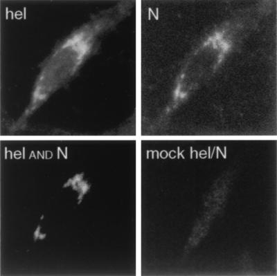
Colocalization of Hel and N in MHV-infected cells. MHV-infected DBT cells were fixed in 100% methanol at 5.5 h p.i. and prepared for confocal immunofluorescence with the anti-N MAb and the anti-Hel rabbit polyclonal serum B1. Hel was imaged at 647 nm (far red), and N was imaged at 488 nm (green). For colocalization (Hel and N), the images were merged with an “and” function requiring the presence of white pixels in both N and Hel images for the white pixels to be seen in the merged image (NIH Image 1.62). Thus, the white pixels in this image were colocalized in the N and Hel images. For the mock Hel-N image, the Hel and N signals were overlapped without filtering, to show the maximum signal from both channels in the merged image.