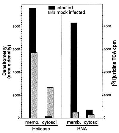
FIG. 8.
Detection of Hel and viral RNA in membrane fractions of MHV-infected cells. MHV-infected DBT cell monolayers were homogenized in the absence of detergent, and differential centrifugation was performed to separate cellular membranes and cytosol, including a final spin at 100,000 rpm with a TLA 20.2 rotor in a Beckman TLX Optima centrifuge. The combined membrane pellets (memb.) and the post-100,000-rpm cytosol were assessed for Hel by immunoprecipitation with the B1 antibody followed by fluorography and densitometric analysis (NIH Image 1.62) with a calculation of area in pixels × the mean density of the pixels. Quantitation of new viral RNA was performed by determination of TCA-precipitable [3H]uridine in the presence of actinomycin D. Mock-infected cells were used as controls.

