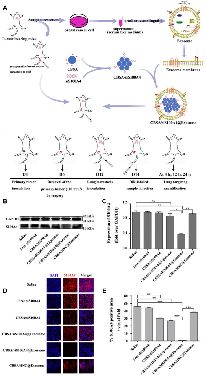
Figure 5.
Schematic representation of postoperative lung metastasis model and drug therapy and S100A4 expression in the lung post-treatment [131]. (A) Schematic illustration of exosome-mediated siRNA delivery to suppress postoperative breast cancer metastasis. (B) Expression of S100A4 in the lung determined by Western blot analysis after treatment with saline, free siS100A4, CBSA/siS100A4, CBSA/siS100A4@Liposome, CBSA/siS100A4@Exosome, and CBSA/siNC@Exosome. (C) S100A4/GAPDH values in the lung tissues of each group; the data represent the mean ± SE (n = 4, * p < 0.05, ** p < 0.01). (D) Immunostaining with anti-S100A4 antibody and Cy3-conjugated secondary antibody (red) showing immunofluorescence images of S100A4 expression in lung tissues from each group. Nuclei were stained with DAPI (blue) and samples were imaged by laser scanning confocal microscopy. Scale bar = 75 μm. (E) Quantitative assessment of S100A4 in treated lung tissue. The data represent the mean ± SE (n = 4, ** p < 0.01, *** p < 0.001).

