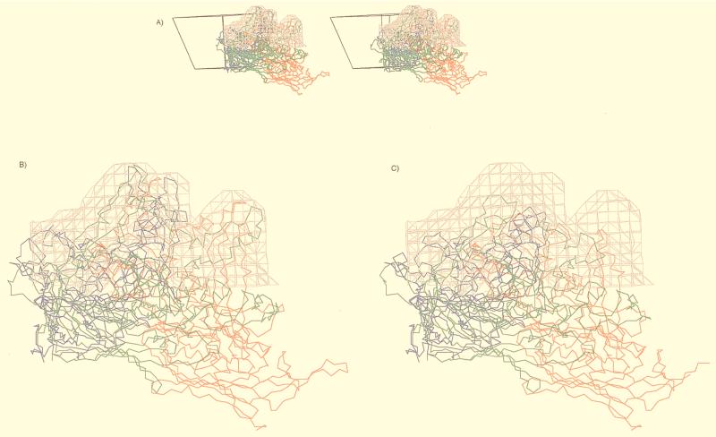
FIG. 6.
The pseudoatomic model of ADV-G VP2 fitted into the cryo-EM reconstruction (gray isodensity contour). (A) Stereo diagram of the mounds viewed parallel to an icosahedral twofold axis, showing the right-hand-side trimer with the reference VP2 in blue and the icosahedral threefold-related VP2s in red and green. Shown also is the viral asymmetric unit depicted as an open triangle. (B) Close-up view of panel A with inserted peptides IN1, IN2, IN4, IN6, and IN7 in ADV. (C) Close-up view of the ADV mound density with superimposed CPV atomic model, which has no equivalence structure to the inserted peptides in ADV. This figure was generated with the program MacInPlot (57).

