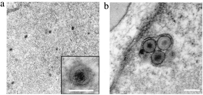
FIG. 4.
Detection of HHV8 particles in infected DMVEC by electron microscopy. (a) View of a DMVEC nucleus showing herpesvirus-like capsid structures. The insert shows an enlarged view of a single virion (bar = 100 nm). (b) View of a cytoplasmic vesicle containing mature virions sectioned within the nuclear plane (bar = 120 nm).

