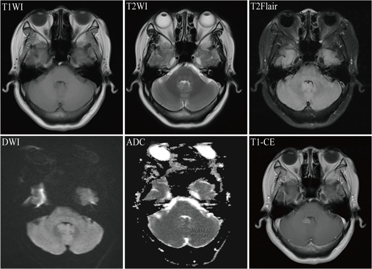
Figure 3.
Magnetic Resonance Imaging (MRI) for breast cancer combined with brain metastasis in a 50-year-old female patient. T1WI revealed an irregular hypointense nodule in the cerebellar vermis; T2WI and T2Flair showed slightly hyperintense signals; diffusion-weighted imaging (DWI) presented an isointense signal; the mean apparent diffusion coefficient (ADC) value of the lesions was 0.813 x 10(-3) mm²/s; and T1-CE demonstrated significant enhancement of the lesion.

