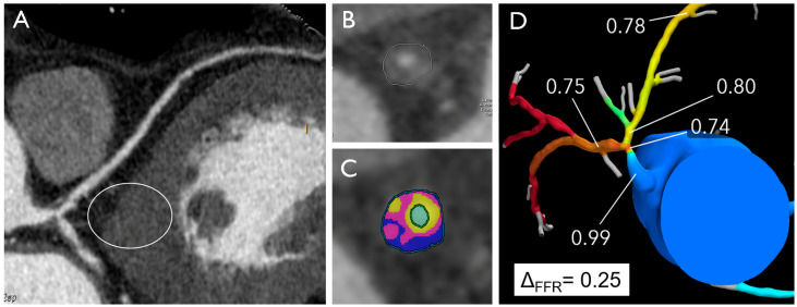
Figure 3.
Distal left main artery plaque in a 53-year-old male with atypical chest pain and normal electrocardiogram. The multiplanar reconstruction image (A) shows a plaque in distal LM with high-risk features (positive remodeling, low attenuation, high plaque volume) (B,C) and positive FFRCT with high delta FFR-CT (D).

