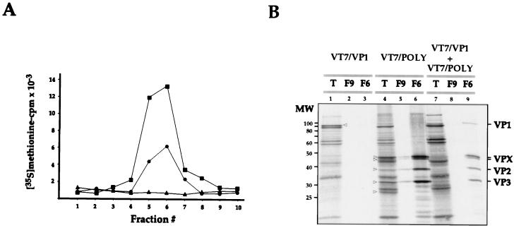
FIG. 2.
Characterization of VLPs produced in cultures coinfected with the rVVs VT7/POLY and VT7/VP1. (A) Cultures infected with VV VT7/POLY (■) or VT7/VP1 (▴) or coinfected with VV VT7/POLY and VT7/VP1 (●) were maintained in medium supplemented with IPTG and rifampin. Cells were metabolically labelled with [35S]methionine. At 24 h p.i., cells were harvested and used to isolate VLPs by velocity sedimentation in sucrose gradients. Gradients were fractionated, and the total amount of acid-precipitable radioactivity was determined by scintillation counting. Fraction 1 corresponds to the bottom part of the gradient. (B) Samples corresponding to the total-cell extract (T) and gradient fractions 9 (F9) and 6 (F6) of samples from cells infected with VT7/VP1 or VT7/POLY or coinfected with VVs VT7/POLY and VT7/VP1 were analyzed by SDS-PAGE and autoradiography. Open arrowheads denote the positions of radioactive bands corresponding to VP1 in lane T from VT7/VP1-infected cells and to the VPX doublet, VP2, VP3, and VP4 in lane T from VT7/POLY-infected cells. The positions of molecular weight markers (MW) and bands corresponding to proteins VP1, VPX, VP2, and VP3 are indicated.

