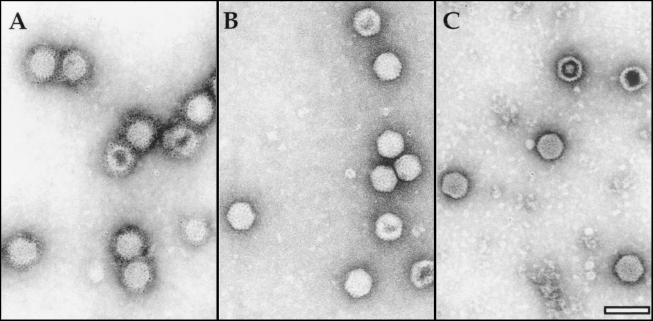
FIG. 4.
Immunoelectron microscopy analysis of VP1-containing VLPs. Purified VP1-containing VLPs were attached to coated EM grids and incubated with rabbit anti-VP2/VPX (A), -VP1 (B), and -VP3 (C), respectively. Bound antibody was detected with goat anti-rabbit Ig conjugated to 5-nm colloidal gold particles. Bar represents 100 nm.

