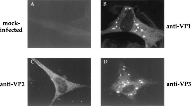
FIG. 6.
Subcellular distribution of IBDV proteins in IBDV-infected CEF. At 24 h p.i., cells were fixed and incubated with rabbit anti-VP1 (anti-VP1), -VP2/VPX (anti-VP2), or -VP3 (anti-VP3) antisera. Thereafter, samples were incubated with FITC-conjugated goat anti-rabbit Ig and viewed by epifluorescence.

