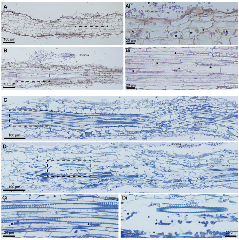
Figure 4.
The histological analysis of B. distachyon roots infected with N. crassa. (A,B) Paraffin sections, 7 μm, stained with hematoxylin and eosin. Conidia outside the root (A,B). Hyphae (*) seen inside a few plant root cortical cells whereas the majority of root cortical cells do not appear to be infected (A). Hyphae (*) growing through the vascular bundle (B). (C,D) Epon sections, 1 μm, stained with Richardson’s solution. Hyphae (*) growing inside the vascular bundle of a moderately infected root with vascular elements preserved (C), and of a strongly infected root with disrupted vascular elements (D). Xylem elements can be recognized by secondary wall thickenings. Panels (i) display details of boxed areas. Asterisk (*): hypha.

