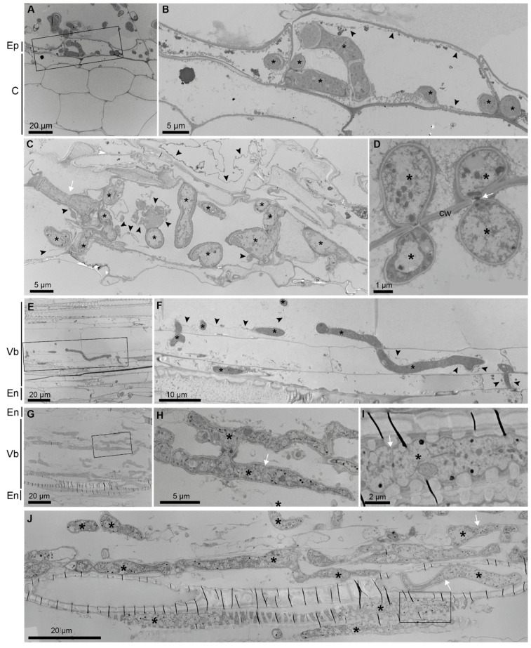
Figure 5.
The TEM analysis of B. distachyon roots infected with N. crassa. Hyphae in epidermal cells (A,B), in cortical cells (C,D), in a moderately infected vascular bundle (E,F), and in a strongly infected vascular bundle (G–J). The plant cell cytoplasm/membrane (arrowheads) can be observed in infected cells and neighboring cells (C). Hyphae spread through the root by crossing plant cell walls (D,F). In a moderately infected root, plant vascular elements are largely preserved (E,F). In a strongly infected root, vascular elements are destroyed except for the xylem elements with secondary wall thickenings (G,J). Parallel, thin, black lines represent folds that were generated over these rigid cell walls during sectioning (G,J). Hyphae grow unrestricted (H) or inside the xylem vascular elements (I,J). The characteristic ultrastructure of fungal hyphae (*) includes the cell wall, septa (with pores) (arrows), and dense cytoplasm. Boxed areas (A,E,G,J) are shown as enlarged (B,F,H,I). Asterisk (*): hypha; white arrow: hyphal septum; black arrowhead: plant cell cytoplasm/membrane; Ep: epidermis; C: cortex; En: endodermis; Vb: vascular bundle; Cw: cell wall.

