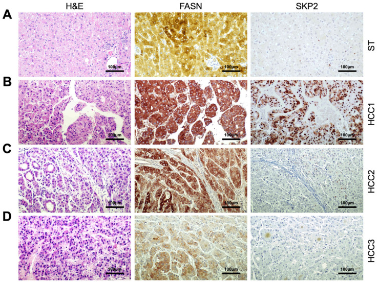
Figure 5.
Representative immunohistochemistry patterns of FASN and SKP2 proteins in human hepatocellular carcinoma (HCC; n = 210). (A) Example of a liver non-tumorous surrounding tissue exhibiting moderate membranous and cytoplasmic for FASN and weak/absent immunolabeling for SKP2. (B) A human HCC (denominated HCC1) displaying robust and diffuse immunoreactivity for FASN and SKP2 proteins. Note that FASN staining is localized in the cytoplasm of HCC cells, whereas SKP2 immunoreactivity is localized in the cytoplasmic and nuclear compartments. (C) A second hepatocellular tumor (HCC2) is characterized by intense, homogeneous FASN immunoreactivity and low/absent SKP2 immunolabeling. (D) Finally, a third tumor (HCC3) shows faint FASN positivity and absent SKP2 immunoreactivity. Abbreviation: H&E, hematoxylin and eosin staining. Original magnifications: 200× in all panels. Scale bar: 100 µm in all panels.

