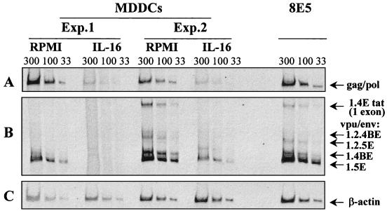
FIG. 1.
Viral mRNA expression in HIV-1Ba-L-infected MDDCs cultured for 12 days in the absence or presence of rIL-16 (3 μg/ml). In two separate experiments (Exp.), RNA samples (33, 100, and 300 ng) were subjected to RT-PCR amplifications with primer pair GAG06-GAG04 to detect Gag or Pol unspliced mRNA (A) and with primer pair BSS-KPNA to detect intermediate-size singly spliced viral transcripts (B). These mRNAs were named according to the exons they contain and the proteins they produce (19): 1.4E Tat (exons 1 and 4E), 1.2.4BE Vpu/Env (exons 1, 2, and 4BE), 1.2.5E Vpu/Env (exons 1, 2, and 5E), 1.4BE Vpu/Env (exons 1 and 4BE), and 1.5E Vpu/Env (exons 1 and 5E). Constitutively expressed β-actin mRNA in the same samples was amplified (C). An equivalent amount of RNA from the 8E5 cell line was used as a positive control of the RT-PCR amplification. RT-PCR products were resolved on a 4 to 5% nondenaturing polyacrylamide gel, visualized under UV light after ethidium bromide staining, and photographed.

