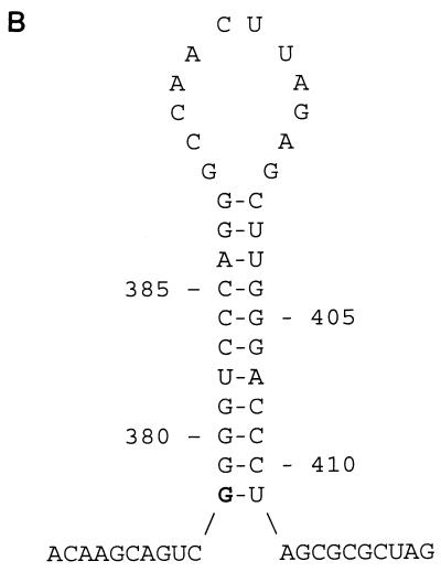
FIG. 2.
(A) Nucleotide sequence alignment of the 5′ and 3′ LTRs of PcEV. Identical nucleotides are indicated by dashes; gaps introduced for optimal alignment are indicated by dots. The LTRs of PcEV differ in length due to the deletion of a direct repeat sequence in the 3′ LTR. The boundaries between U3, R, and U5 are indicated by vertical lines and arrows. The four GATA-1 binding sites ([A/T]GATA[A/G]), the CAAT box (CCAAT), the TATA box (TATATAA), the polyadenylation site (AATAAA), and the flanking indirect repeats (IR) are indicated by bold letters. The direct repeat sequences DR1A to DR1E are indicated by underlining and demarcated by vertical lines. (B) Predicted RNA secondary structure at the 5′ end of the R region in the LTRs of PcEV. The length of the stem is 11 nt, the size of the loop is 12 nt, and the free energy value (at 25°C) is −20.6 kcal. The predicted first nucleotide of viral RNA is indicated by bold type.


