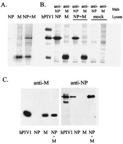
FIG. 1.
Expression of the hPIV-1 M and NP genes in 293T cells with the mammalian expression vector pCAGGS. (A) The culture media were clarified, and cellular materials released into the culture media were concentrated by pelleting them through a cushion of 50% glycerol–phosphate-buffered saline. The pellets were resuspended in Laemmli reducing sample buffer, and equal aliquots of all samples were analyzed by SDS-PA gel electrophoresis (9% PA gel). Culture supernatants of cells transfected with the hPIV-1 NP gene alone, the hPIV-1 M gene alone, and the hPIV-1 NP and M genes together are shown. (B) Cell lysates were immunoprecipitated with monoclonal antibodies (MAb) against hPIV-1 M (P3B) or hPIV-1 NP (P27), and immunoprecipitates were resolved in a 9% PA gel. hPIV-1, purified hPIV-1 as a marker. (C) Western blot analysis of proteins released from transfected cells into media. The same samples shown in panel A were transferred to nitrocellulose membranes and reacted with rabbit polyclonal antibody against SDS-denatured hPIV-1 M protein (left column) or with monoclonal antibody against hPIV-1 NP (P27). hPIV-1, 1 μg of purified hPIV-1 used as control.

