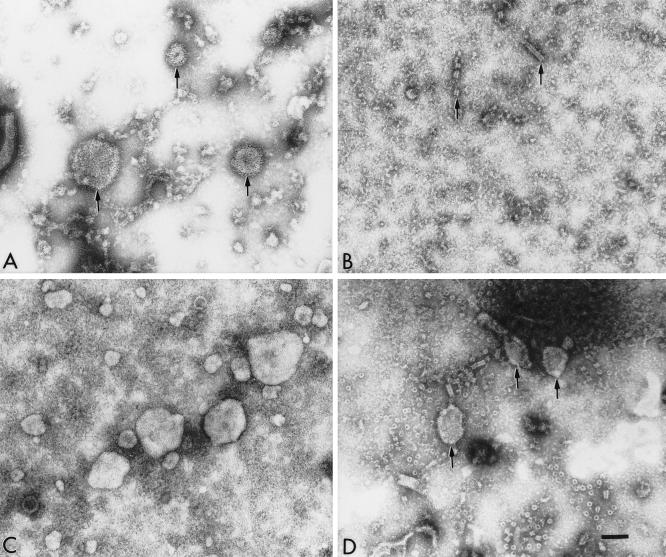
FIG. 2.
Electron micrographs of the components observed in the culture media of cells infected with hPIV-1 (A) or cells expressing the hPIV-1 NP gene (B), the hPIV-1 M gene (C), or the hPIV-1 M and NP genes (D) from pCAGGS plasmids. The clarified culture media of 293T transfected cells were concentrated in Microcons 100 (Amicon) by centrifuging them at 1,000 × g for 25 min. Concentrated media were adsorbed to freshly glow-discharged, carbon-coated grids, negatively stained with 2% phosphotungstic acid, and observed in a Phillips 301 electron microscope operated at 60 kV. The bar represents 100 nm. (A) hPIV-1 virions in culture medium (arrows); (B) occasional NC-like structures (arrows) observed in the culture medium from cells expressing the NP gene; (C) medium from cells expressing the hPIV-1 M gene; (D) medium from cells coexpressing the hPIV-1 M and NP genes (arrows).

