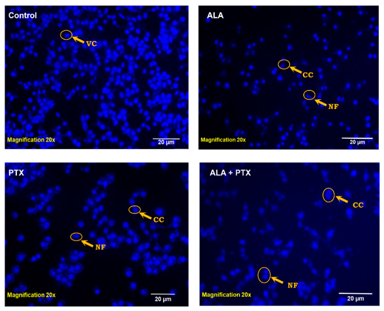
Figure 7.
The DAPI-stained nuclei of MCF-7 cells are shown in the figure with changes in their morphology. Furthermore, DAPI staining following treatment with PTX-Lipo, ALA-Lipo, and ALA-PTX-Lipo demonstrated VC (viable cells), CC (chromatin condensation), and NF (nuclear fragmentation), indicated by an arrow, in comparison to the control group.

