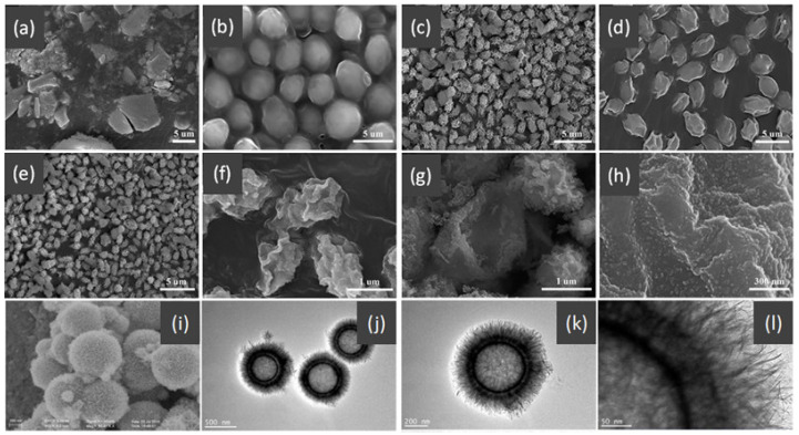
Figure 18.
SEM images of the (a) Fe-doped CeO2 nanoparticles, (b) yeast template, (c) CeO2 hollow microspheres, and Fe-doped CeO2 hollow microspheres before (d) and after (e–h) calcination. Figure reproduced from ref. [211]. The SEM image (i) and TEM images (j–l) of Fe3O4@MnO2 BBHs. Figure reproduced from ref. [212].

