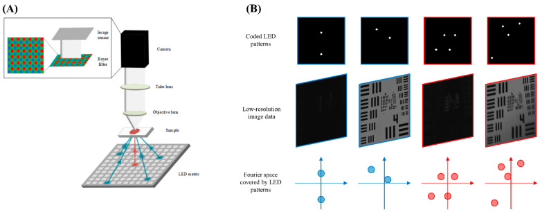
Figure 1.
Fundamentals of FPM microscope technology. (A) Schematic of experimental FPM microscope demonstrates the microscope using conventional one-LED illumination (red arrow) and multiplexed four-LED illumination (blue arrows). (B) Samples of multiplexed illumination patterns. Samples represent two-LED (blue) and four-LED (red) illumination patterns. The top row of images represents positions of activated LEDs on the grid for each image. The middle row shows the raw low-resolution images captured using the activated LEDs. The bottom row illustrates the Fourier space subregions sampled by the captured raw images.

