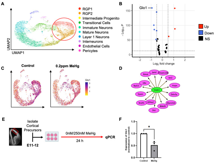
Figure 1.
Glo1 expression is reduced in radial glial precursors (RGPs) following prenatal low-dose methylmercury (MeHg) treatment. (A) Visualization of cells from control (0 ppm) and MeHg (0.2 ppm) treated cortical tissue, colored by Seurat clustering and annotated by cell type, red circle represents cell populations (RGP1 and RGP2) used for downstream differentially expressed gene (DEG) analysis (B) Volcano plot of differentially expressed genes between control (0 ppm) RGP1 and RGP2 and MeHg (0.2 ppm) RGP1 and RGP2. Discriminated based on p-value adjusted and log2 fold-change. Log2 fold-change > 0.5 and p-value adjusted < 10e−14. (C) Visualization of the total cell population after PCA and UMAP, colored by expression of Glo1. (D) Transcription factor CREB1 was identified from iRegulon (Cytoscape) and its direct transcriptional targets. (E) Experimental timeline following radial glia precursor (RGP) isolation from embryonic day 11–12 (E11–12) CD1 mice, created with BioRender.com. The red box indicates the region dissected to obtain RGPs. (F) Cells were exposed to two conditions: (i) control (0 nM MeHg) and (ii) 250 nM MeHg for 24 h, at which point they were lysed. Quantitative analysis of Glo1 expression, over GAPDH, normalized to control. n = 4 independent experiments, Student t-test, * p < 0.05. Error bars indicate the SEM.

