
Figure 16.
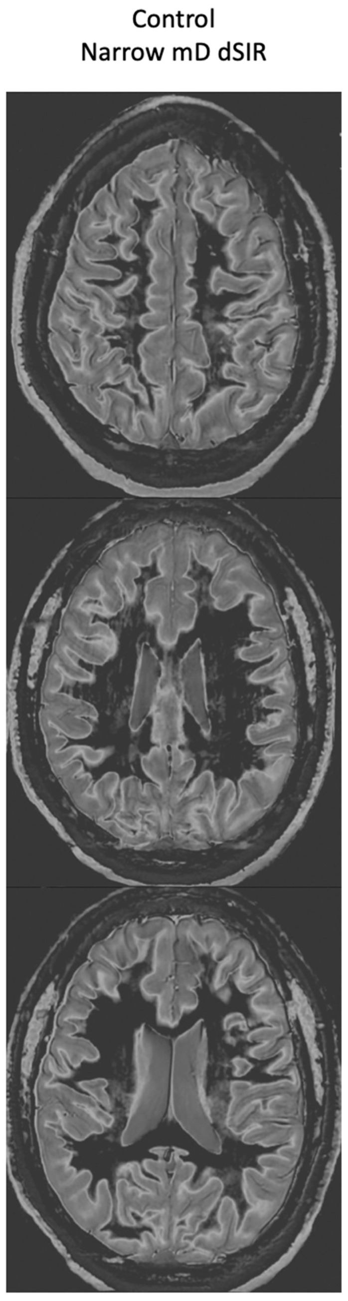
Normal 18-year-old control. 2D narrow mD dSIR images. The narrow mD dSIR images show normal white matter as very low signal intensity (dark) except for intermediate areas in and around the corticospinal tracts. This is a whiteout sign grade 1. Normal high signal boundaries are seen at the junction between white matter and gray matter as well at the junction between white matter and CSF around the lateral ventricles.