Figure 1.
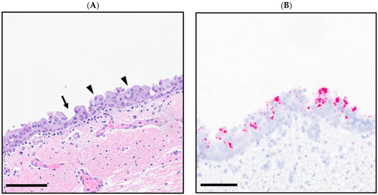
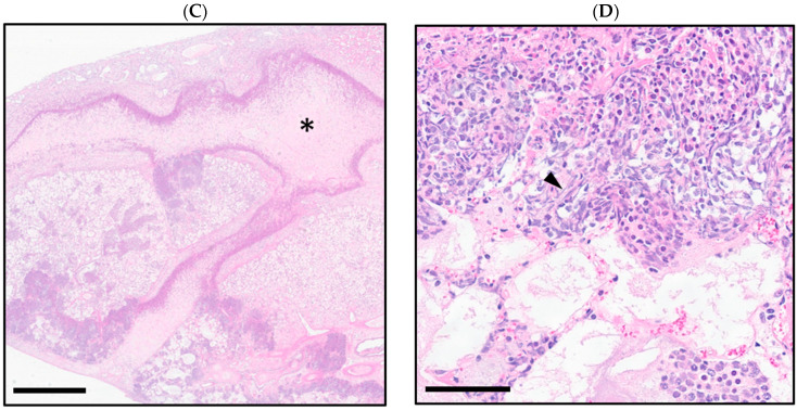
(A) BRBV variant-infected goat bronchial epithelium with antibasilar nuclei, loss of cilia (arrowheads), and segmental attenuation (arrow); (hematoxylin and eosin-stained; scale bar = 80 µm). (B) BRBV variant-infected goat bronchus; RNAscope in situ hybridization for BRBV variant (red; hematoxylin counterstain; scale bar = 60 µm). (C) Goat lung with multifocal, mosaic pattern of coagulative necrosis surrounded by degenerate leukocytes (asterisk) (hematoxylin and eosin-stained; scale bar = 1 mm). (D) Goat lung in (C), highlighting the presence of degenerate leukocytes with nuclear streaming (arrowhead) surrounding regions of necrosis (hematoxylin and eosin-stained; scale bar = 60 µm).


