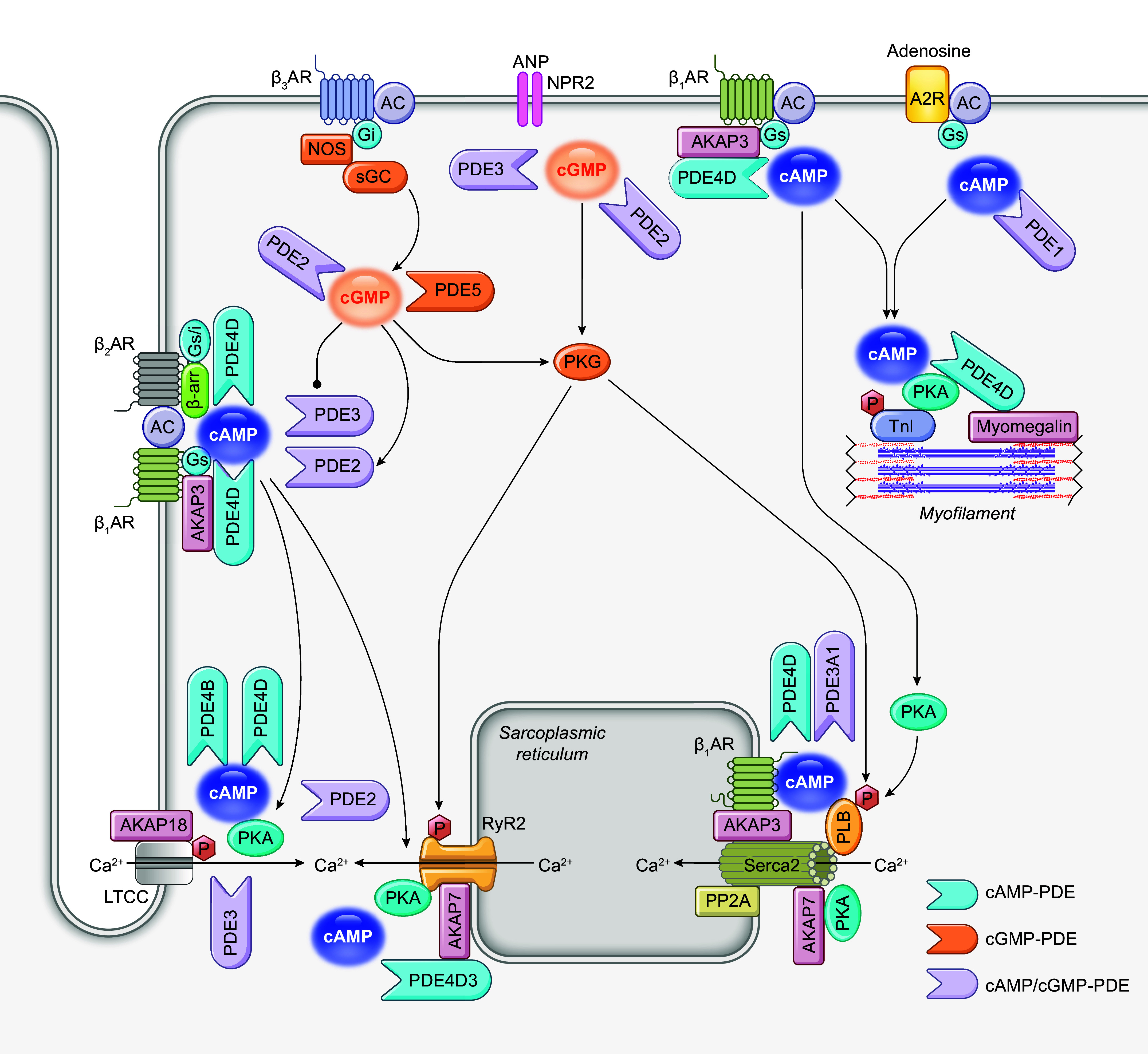
FIGURE 5.

Schematic illustration of phosphodiesterases (PDEs) in cardiac excitation-contraction coupling. A2R, adenosine receptor 2, AC, adenylyl cyclase; ANP, atrial natriuretic peptide; AKAP, A-kinase anchoring protein; β-AR, β adrenergic receptor; cAMP, cyclic adenosine monophosphate, cGMP, cyclic guanosine monophosphate; Gi, inhibitor G protein; Gs, stimulatory G protein; LTCC, L-type calcium channel; NPR2, natriuretic peptide receptor 2; PKA, protein kinase A; PKG, protein kinase G; PLB, phospholamban; PP2A, protein phosphatase 2A; RyR2, ryanodine receptor 2; SERCA, sarco(endo)plasmic reticulum calcium ATPase; sGC, soluble guanylyl cyclase; TnI, troponin I.
