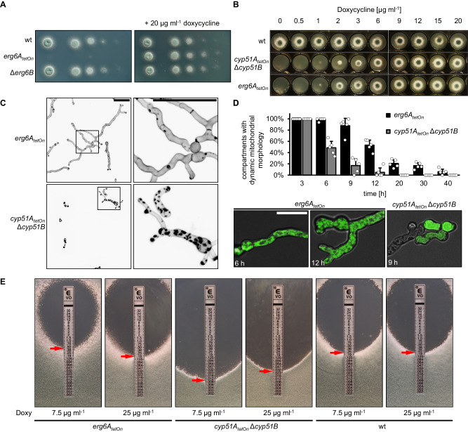
Fig. 2. The impact of sterol C24-methyltransferase depletion on viability, cell wall patch formation, and azole susceptibility.
A In a series of tenfold dilutions derived from a starting suspension of 5 × 10⁷ conidia ml−1 of wild type (wt), the conditional mutant erg6AtetOn, and the deletion mutant Δerg6B, aliquots of 3 µl were spotted onto AMM agar plates. AMM was supplemented with 20 μg ml−1 doxycycline when indicated. Agar plates were incubated at 37 °C, representative photos were taken after 28 h. B 1.5 × 10³ conidia of the indicated strains were spotted on AMM agar supplemented with the indicated amount of doxycycline. The plates were then incubated at 37 °C, representative photos were taken after 40 h. C, D Conidia of the erg6AtetOn and cyp51AtetOn Δcyp51B mutants which express mitochondria-targeted GFP (D) or not (C) were inoculated in Sabouraud medium supplemented with 20 µg ml−1 doxycycline and incubated at 37 °C. After 6 h, doxycycline was depleted by washing the wells three times with Sabouraud medium without doxycycline. The hyphae were then incubated in Sabouraud medium without doxycycline at 37 °C for an additional 40 h. C Hyphae were stained with calcofluor white and analyzed with a confocal microscope. Depicted are representative images of z-stack projections of optical stacks of the calcofluor white fluorescence covering the entire hyphae in focus. The right images show magnifications of the framed sections in the left images. Bars represent 50 μm and are applicable to all images in the respective panel. Data were representative of five independent experiments. (D, column graph) At the indicated time points after doxycycline depletion, the viability of the hyphae of the indicated strains expressing mitochondria-targeted GFP was analyzed with time-lapse spinning disc confocal microscopy. The bars indicate the percentage of hyphal compartments with evident mitochondrial dynamics (viable compartments). Each bar represents the mean of five technical replicates (data points) for each timepoint, with an average of ~80 analyzed compartments per strain for each timepoint. The error bars indicate standard deviations based on the five technical replicates. (D, lower panel) Representative overlay images of the bright-field channel and of z-stack projections of optical stacks of the GFP fluorescence covering the entire hyphae in focus. Depicted are exemplary images of a viable hypha with tubular mitochondrial morphology (also showing mitochondrial dynamics in time-lapse microscopy; left image) and of hyphae with fragmented mitochondria (showing no mitochondrial dynamics in time-lapse microscopy; middle image) or with no or cytosolic GFP signal (right image) which were considered to be dead. Bars represent 50 μm and are applicable to all respective subpanels. E 1 × 10⁶ conidia of the indicated strains were spread on Sabouraud agar plates. Agar was supplemented with the indicated amount of doxycycline (Doxy) to achieve a different induction of the conditional promoters. Voriconazole Etest strips were applied. The plates were incubated at 37 °C and representative photos were taken after 42 h.

