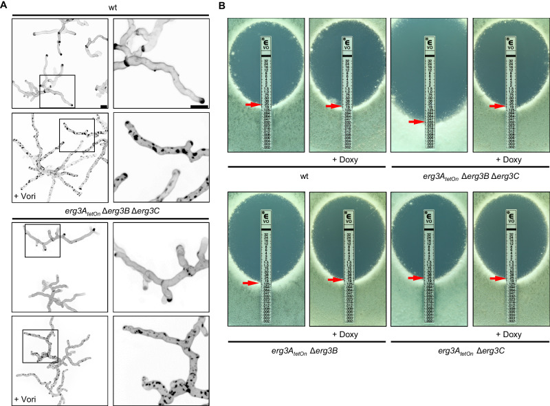
Fig. 6. The influence of the sterol C5-desaturase on azole susceptibility and azole-induced cell wall patch formation.
A Conidia of wild type and the conditional sterol C5-desaturase mutant (erg3AtetOn Δerg3B Δerg3C) were inoculated in the Sabouraud medium. After 10 h incubation at 37 °C, the samples were either directly analyzed (controls without azole) or the medium was supplemented with 4 µg ml−1 voriconazole (+Vori). The voriconazole-exposed hyphae were then incubated at 37 °C for another 5 h. The untreated hyphae (upper images per indicated strain) and the voriconazole-treated hyphae (lower images per indicated strain) were stained with calcofluor white and analyzed with a confocal microscope. Depicted are representative images of z-stack projections of optical stacks of the calcofluor white fluorescence covering the entire hyphae in focus. The right panels show magnifications of the framed sections in the left panels. Bars represent 50 μm and are applicable to all images in the respective panel. Data were representative of five independent experiments. B 1 × 10⁶ conidia of wild type (wt) and the indicated strains were spread on Sabouraud agar plates. When indicated, agar was supplemented with 25 μg ml−1 doxycycline (+Doxy). Voriconazole Etest strips were applied. The plates were incubated at 37 °C and representative photos were taken after 42 h.

