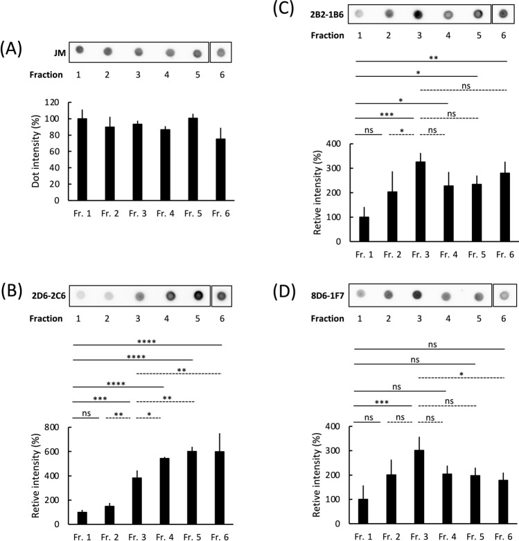
Figure 1.
Binding of antibodies against granular tau oligomers to fractionated recombinant tau aggregates. Recombinant human full-length (FL) tau was polymerized by heparin and then fractionated into Fractions (Fr) 1–6 through sucrose step gradient centrifugation. As the fraction number increases, denser tau aggregates are observed 19. For instance, Fr. 1 contains monomeric and multimeric tau, while granular tau oligomers are predominantly found in Fr. 3. Fibrils are present in Fr. 4–6. Dot blot analysis detected tau in Fr. 1–6 using the pan-tau rabbit polyclonal antibody JM (A) and monoclonal antibodies such as 2D6-2C6 (B), 2B2-1B6 (C) and 8D6-1F7 (D), derived from rat immunized with granular tau oligomers. Fraction 1 contained tau at a concentration of 100 μg/ml. Tau immunoreactivity was quantified by densitometry (ImageJ software), and the levels of 2D6-2C6, 2B2-1B6 and 8D6-1F7-reactive tau were normalized to the corresponding JM-reactive tau. Quantitative data are presented as a percentage of Fr. 1 (mean ± SD from 4 experiments). P values were determined using one-way ANOVA followed by Tukey’s multiple comparisons test. Significance levels are indicated as *(p < 0.05), **(p < 0.01), ***(p < 0.001), ****(p < 0.0001), and ns denotes not significant. There are annotations on solid lines (comparing Fr. 1 to Fr. 2, 3, 4, 5 or 6) and dotted lines (comparing Fr. 3 to Fr. 2, 4, 5 or 6). The dotted signals of Fr. 6 were cropped from different parts of the same membrane (Supplemental Fig. 5A–D).

