Figure 3.
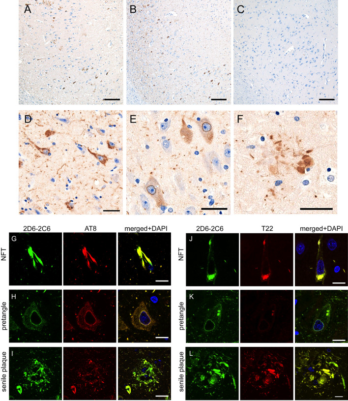
Immunohistochemistry and double immunofluorescence using 2D6-2C6 antibody on autopsied AD cases. (A–F) Immunohistochemistry using 2D6-2C6 antibody on autopsied AD cases. The panels (A–C) display CA1 of autopsied samples. Two cases with AD (A, B) demonstrated 2D6-2C6-immunopositivity in the CA1, whereas a neurologically healthy control (C) did not show dense aggregates. The 2D6-2C6 antibody immunolabeled NFTs (D), pretangles (E), and neuritic plaque (F) in the CA1. Black scale bars = 100 μm (A–C) and 20 μm (D–F). (G–L) Double immunofluorescence using 2D6-2C6 antibody combined with other anti-tau antibodies. All panels here are taken at the CA1 from the same AD case. Panels (G–I) indicate a combination of 2D6-2C6 and AT8, whereas panels (J–L) indicate a combination of 2D6-2C6 and T22. The 2D6-2C6 fluorescent signals were mostly merged with AT8 signals for NFT (G), pretangle (H), and neuritic plaque (I). The 2D6-2C6 and T22 antibodies also exhibited colocalization (J–L). Pretangle (K) was more sensitively detected by 2D6-2C6 antibody compared to T22. White scale bars = 10 μm.

