Figure 5.
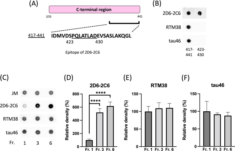
Binding of other antibodies against C-terminal regions with tau aggregates. (A) The sequence at amino acid residues 417–441 in the tau C-terminal region is illustrated. The epitope of 2D6-2C6 covers residues 423–430. (B) Peptides (417–441 and 423–430) were dotted onto a nitrocellulose membrane and probed with the 2D6-2C6 antibody, as well as other C-terminal monoclonal antibodies: RTM38 (immunogen: 417–441) and tau46 (epitope: 404–441). (C–F) Recombinant human full-length tau was polymerized using heparin and then fractionated. Monomer and multimer tau, granule tau oligomers, and longer fibrils are observed in Fraction 1, 3, and 6, respectively. A dot blot analysis detected tau in all three fractions using the pan-tau rabbit polyclonal antibody (JM), 2D6-2C6, RTM38, and tau46 (C). Tau immunoreactivity was quantified by densitometry (using ImageJ software). Levels of 2D6-2C6 (D), RTM38 (E), and tau46 (F)-reactive tau were normalized by the corresponding JM-reactive tau levels. Quantitative data are presented as a percentage of Fraction 1 (mean ± SD of 4 experiments). P values were determined using one-way ANOVA followed by Tukey’s multiple comparisons test. **** indicates p < 0.0001.

