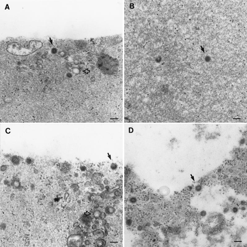
FIG. 5.
Analysis of coupled transcription and translation reactions by thin-section electron microscopy. M-PMV gag genes were expressed in vitro, the reaction mixtures were diluted and centrifuged at high speed, and the resulting pellets were fixed and processed for microscopy. (A to D) Pellets derived from lysates expressing M100A (wild type) Δ26-53, Δ54-83, and R55W, respectively. Solid arrows indicate apparently completed immature capsid structures. Open arrows indicate vesicle-like structures in these non-detergent-treated samples.

