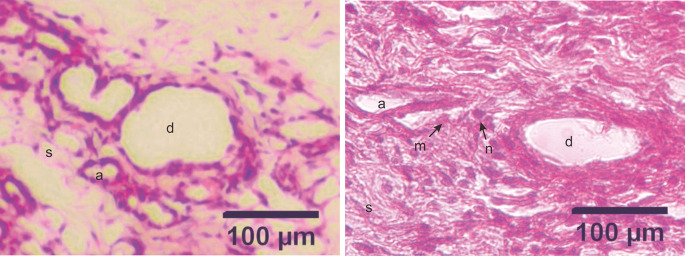
Figure 3. Micrograph of alveoli and mammary ducts in mammary tissue from control group rats and DMBA treatment group rats, with hematoxylin and eosin staining.
A. Control group. The alveoli contain irregular and cuboid cells with no hyperchromatic nuclei. B. Treatment group. The alveoli in the treatment group showed an irregular and indistinct arrangement. The epithelial cells were not cuboidal and contained hyperchromatic nuclei. The mammary ducts were filled with proliferating epithelial cells from the surface of the ducts to the lumen of the ducts. (a) alveoli; (d) ducts; (s) stroma; (m) malignant cells (the cells appeared larger); (n) nuclei (hyperchromatic with a large size). Scale bar, 100 μm. DMBA, 7,12-dimethylbenz[a]anthracene.

