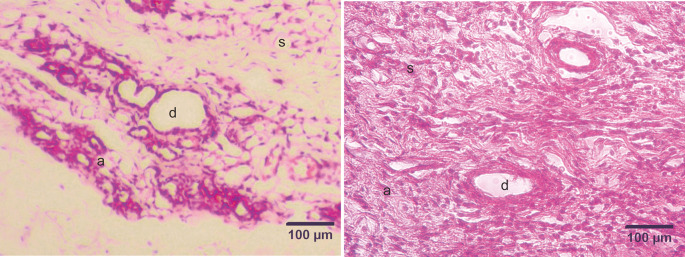
Figure 2. Micrograph of the mammary stroma in mammary tissue from control group rats and DMBA treatment group rats, with hematoxylin and eosin staining.
A. Control group. B. Treatment group. The mammary stroma showed the formation of new epithelial cells that were cancer cells and spread to surrounding tissues and hyperplasia with mitotic fibrocytes in the dermis. (a) alveoli; (d) ducts; (s) stroma. Scale bar, 100 μm. DMBA, 7,12-dimethylbenz [a]anthracene.

