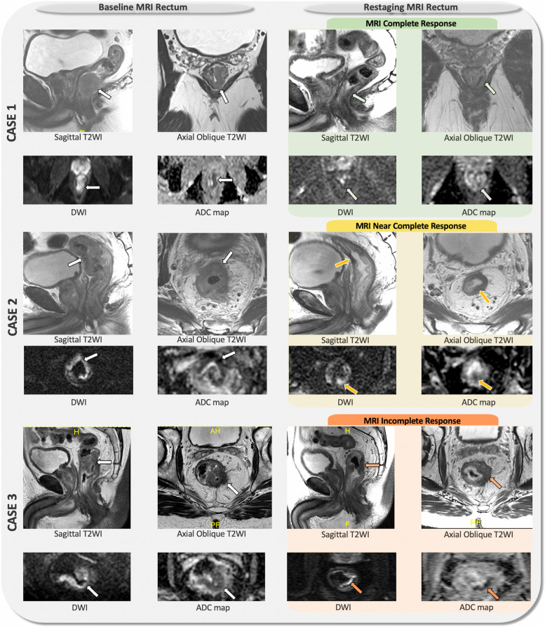
Fig. 3.
Baseline and re-staging MRI of the rectum of patients with complete response (case 1), near complete response (case 2) and incomplete response (case 3) based on the MRI assessment is shown. The white arrows on baseline MRI rectum indicate the primary rectal tumor, which shows intermediate signal intensity (SI) on T2-weighted imaging (T2WI), with high SI on diffusion-weighted imaging (DWI), and low SI on the ADC map, indicating restricted diffusion within the primary tumor. Re-staging MRI on case 1 shows clinical complete response (green box) which is characterized by normalized rectal wall or only fibrosis (low signal intensity on T2WI, green arrows on T2WI) and no areas of restricted diffusion on DWI and ADC map (green arrows on DWI and ADC map). Re-staging MRI on case 2 shows near complete response (yellow box) defined as small area of intermediate SI on T2WI within the tumor bed fibrosis (yellow arrows on T2WI) and small areas of restricted diffusion (yellow arrows on DWI and ADC map). Re-staging MRI on case 3 shows incomplete response (orange box) characterized as significant areas of viable tumor with intermediate SI on T2WI within the tumor bed (orange arrows on T2WI) with restricted diffusion (orange arrows on DWI and ADC map). Reviewing the baseline MRI is highly recommended, as it helps to localize the tumor bed and guide the MRI to the appropriate angulation for high-resolution axial oblique T2WI acquisition perpendicular to the tumor bed

