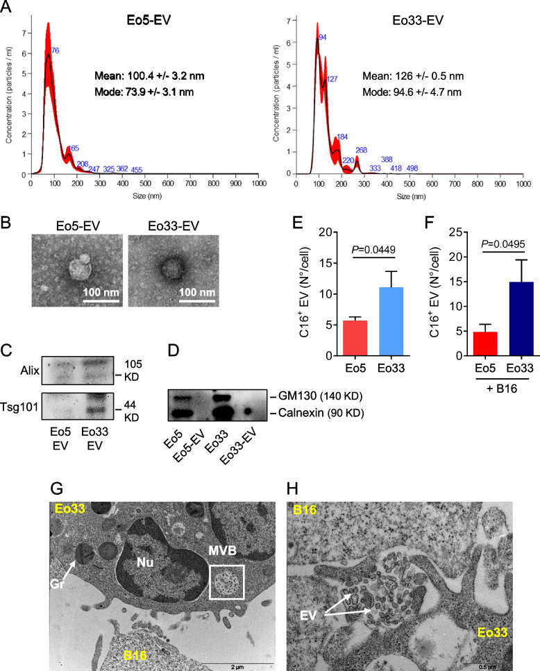
Fig. 1.
IL-33 stimulates the release of EV by eosinophils. EV were isolated from Eo5 and Eo33 conditioned media by serial ultracentrifugations. A NTA analysis of vesicle size and (B) transmission electron microscopy of negative stained Eo5-EV and Eo33-EV. Western blotting for (C) EV markers Alix and Tsg101 in Eo5-EV and Eo33-EV and (D) GM130 and Calnexin in Eo5-EV, Eo33-EV and their producing cells. Flow cytometry quantification of fluorescent EV released by Bodipy FL-C16 labelled Eo5 and Eo33 (E) cultured alone or (F) in the presence of B16.F10 melanoma cells (20:1 ratio) for 24 h. Data are expressed as number of EV released per cell. Mean (SD) of four (E) and three (F) separate experiments is shown. G-H Transmission electron microscopy analysis of Eo33 co-cultured with B16.F10 melanoma cells (10:1 ratio) for 1 h, showing the presence of multivesicular bodies (MVB, G) and EV release (H) by an eosinophil in proximity of a tumor cell. Gr: granule. Nu: nucleus

