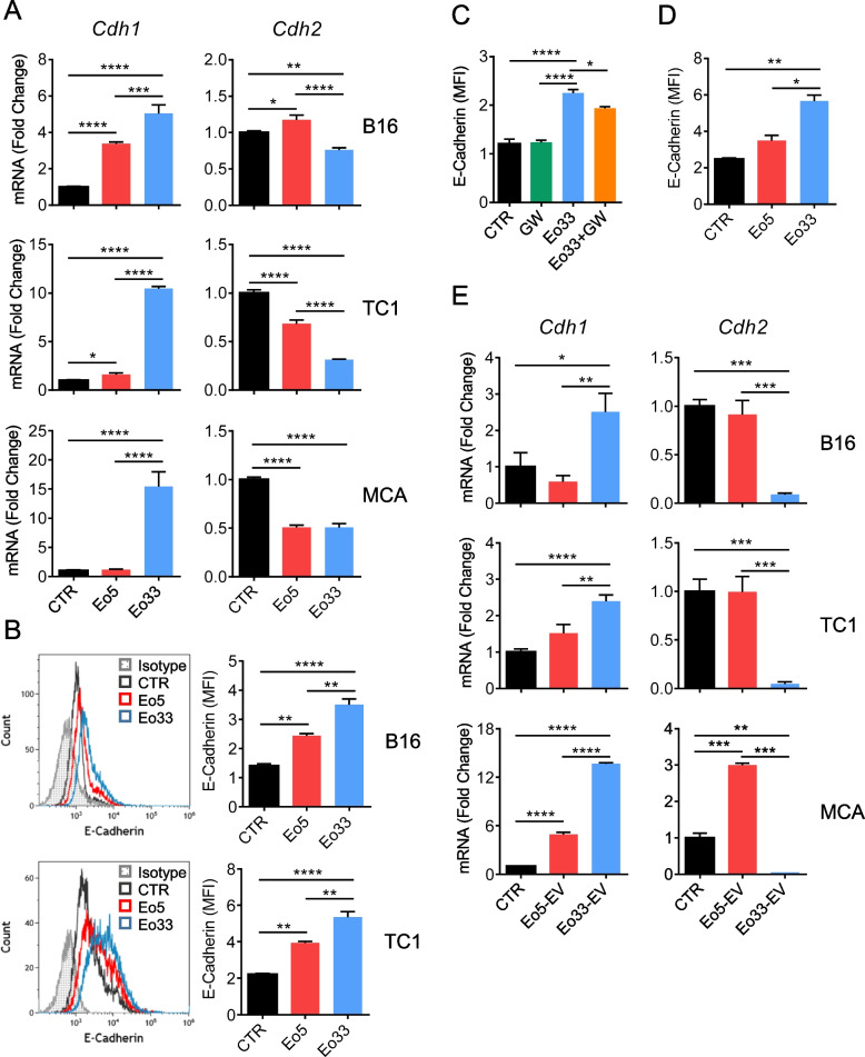
Fig. 4.
IL-33 activated eosinophils regulate EMT markers through EV. Tumor cells were cultured for 24 h alone (CTR) or in the presence of Eo5 or Eo33 (4:1 Eo:Tum ratio). A Expression of CDH1 (E-Cadherin) and CDH2 (N-Cadherin) genes in the indicated cell lines. Mean (SD) of three replicates is shown. *P < 0.05; **P < 0.01; ***P < 0.001; ****P < 0.0001. B Flow cytometry analysis of the expression levels of E-Cadherin on tumor cells. Left, representative histograms. Right, mean fluorescence intensity (MFI) of three replicates, mean (SD). **P < 0.01; ****P < 0.0001. E-Cadherin expression on B16 melanoma cells (C) following 24 h co-culture with GW4869 pre-treated or untreated Eo33 or (D) cultured with Eo33 in a 0.4 μm Transwell system (24 h). Mean (SD) of three replicates is shown. *P < 0.05; **P < 0.01; ****P < 0.0001. E Expression of CDH1 and CDH2 in the indicated tumor cell lines cultured for 24 h alone (CTR) or in the presence of Eo5-EV or Eo33-EV. Mean (SD) of three replicates is shown. *P < 0.05; **P < 0.01; ***P < 0.001; ****P < 0.0001

