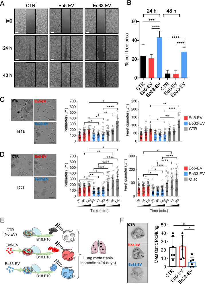
Fig. 5.
Phenotypic changes in tumor cells following incorporation of Eo33-EV. A Wound-healing scratch assay. B16 melanoma cells were exposed to Eo5-EV, Eo33-EV or left untreated for 24 h and then scratched. Phase-contrast pictures were taken at the indicated times. Scalebar, 100 μm. B Quantification of cell-free area by ImageJ analysis at indicated times. Mean (SD) of several fields is shown. ***P<0.001; ****P < 0.0001. Morphologic changes in (C) B16 melanoma cells and (D) TC1 lung adenocarcinoma cells after exposure to Eo5-EV or Eo33-EV. Representative microphotographs at 140 min of culture (left); scalebar, 100 μm. Feret’s diameter and cell perimeter (right) were extrapolated from time lapse video (1440 min., frame interval 20 min). Histograms represent the mean values (SD) of several cells from several fields. Dots represent single cells. *P < 0.05; **P < 0.01; ***P < 0.001; ****P < 0.0001. E Experimental melanoma pulmonary metastasis assay. B16 melanoma cells were exposed to Eo5-EV, Eo33-EV or left untreated for 24 h and then injected intravenously (2 × 10⁵) in C57Bl/6 mice. Mice were sacrificed 14 days later to examine metastases formation in the lungs. F Representative images of metastatic lungs for each experimental group (left) and lung metastatic foci counts (right). Mean (SD) is shown (n = 6 mice/group). *P < 0.05

