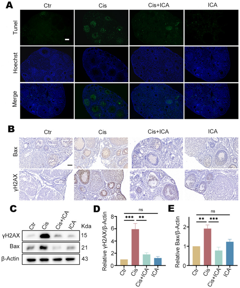
Figure 3.

ICA inhibits cisplatin-induced apoptosis in mouse ovaries. (A) TUNEL was performed on each group’s ovarian sections, and green fluorescent signals showed apoptosis. The nuclei of the cells were stained with Hoechst and fluoresced blue (n = 3). Scale bars, 100 µm. (B), Representative IHC images of Bax and γH2AX in each group of ovaries (n = 6). Scale bars, 100 µm. (C), Detection of γH2AX and Bax protein expression levels in ovarian tissues by WB (n ≥ 3). β-Actin was used as an internal control. (D,E), Quantitative analysis of γH2AX and Bax protein levels. Data are presented as mean ± SEM, **P < 0.01, ***P < 0.001, ns, non-statistically significant.
