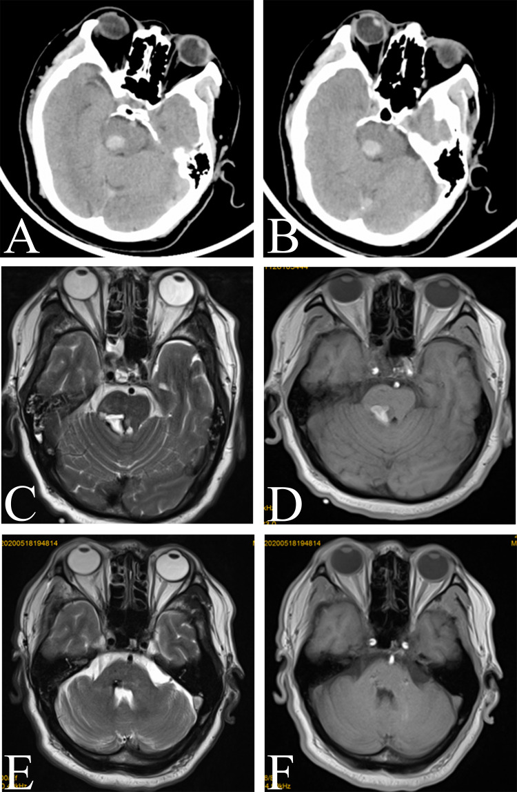
Figure 1.
Image of a right pontine hemorrhage near the red nucleus. (A and B) Preoperative computed tomography image showing acute intracerebral hemorrhage in the right pons near the red nucleus. (C and D) MR image showing that the brainstem hemorrhage was located in the right pontine arm. (E and F) Preoperative T2-weighted and T1-weighted axial magnetic resonance images revealing the resolution of hemorrhage. Encephalomalatic changes, cystic degeneration, and hemosiderin around the lesion were also absorbed.

