
Figure 9. Overexpression of miR-223 in vivo reduces tumor hypoxia and PD-1/PD-L1 expression in DEN+CCl4-induced HCC.
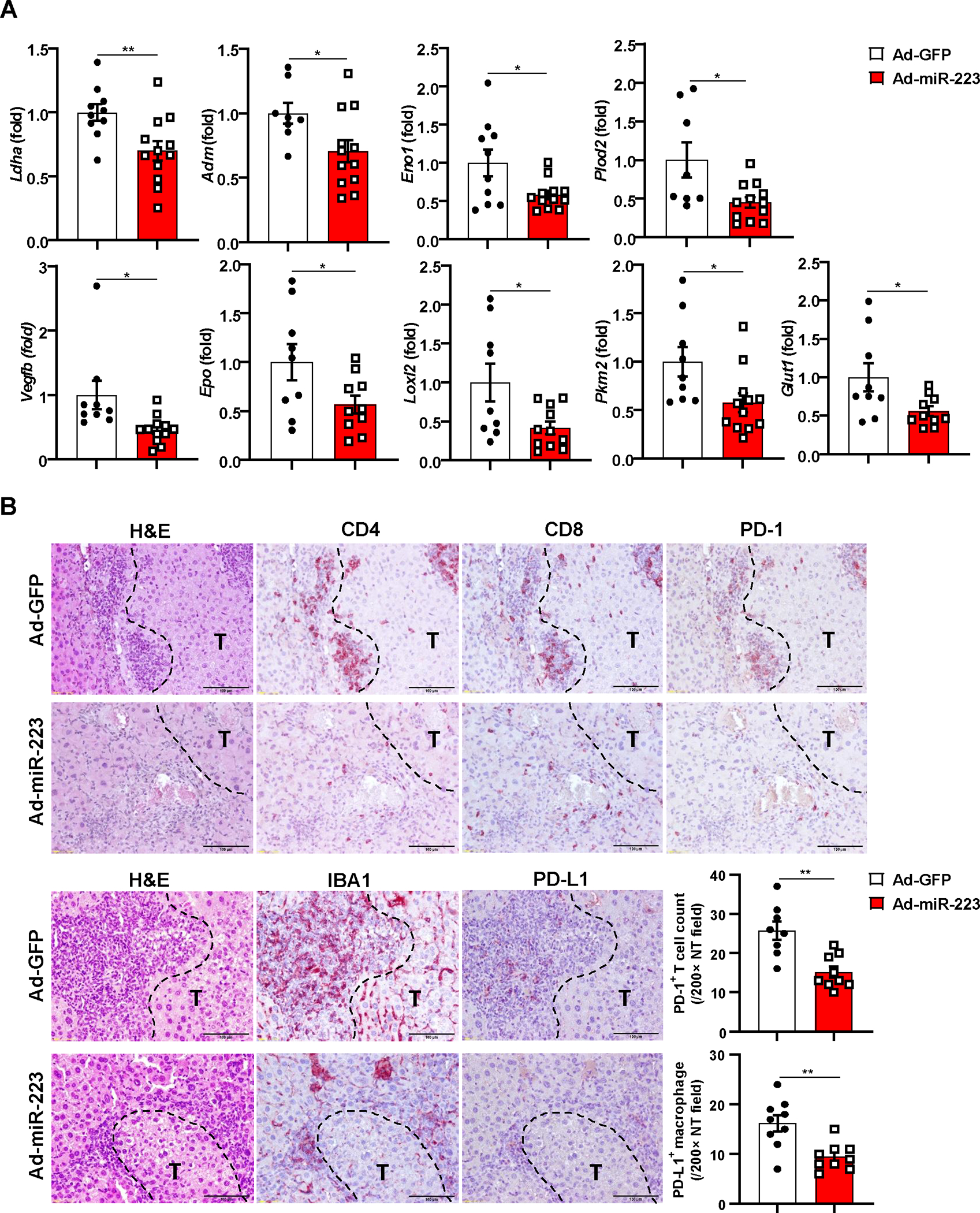
C57BL/6J mice were treated with DEN+CCl4 as described in Figure 1, and were treated with Ad-miR-223 or Ad-GPF. (A) RT-qPCR analyses of HIF-1α target genes in HCC samples from Ad-miR-223 and control Ad-GFP-treated mice. (B) Upper panel: representative immunostaining for infiltrated PD-1+ CD4+/CD8+ T cells surrounding tumor region are shown. Scale bar: 200μm. Lower left panel: representative images for PD-L1+ macrophages surrounding tumor region are shown. Lower right panel: counts of PD-1+ T cells and PD-L1+ macrophages in non-tumor region (NT) were quantified and are shown. Tumor region was labeled with ‘T’; tumor border was depicted with black dash line. Values represent means ± SEM. *P< 0.05, **P< 0.01.